Professor Jin Yuan's team from Zhongshan Ophthalmic Center established a new standard for the diagnosis and grading of meibomian gland dysfunction, published on EClinicalMedicine
Resource: Ophthalmic Engineering Research Center
Written by: Penf Xiao, Ophthalmic Engineering Research Center
Proofread by: Jiawei Wang
Edited by: Xianjing Wei
Reviewed by: Xiaoling Liang
Meibomian Gland Dysfunction (MGD) is one of the most common ocular surface diseases extremely prevalent worldwide, especially in Asian populations (from 46.2% to 69.3%), that degrades patients’ ocular comfort, visual function, and quality of life. As the most common trigger of dry eye diseases (DED), early diagnosis and grading strategy for MGD will optimize treatment algorithms. However, existent diagnostic tests based on the observation of abnormal anatomy and physiology of the MGs opening and lid secretions are subjective, limited in detection of early stage of MGD. Prof. Jin Yuan's team from Zhongshan Ophthalmic Center has developed an objective multi-parametric meibomian glands analyser to quantitatively analyse the meibomian glands using infrared meibography images (Quantitative Imaging in Medicine and Surgery, 11(4), 2021). A real world study aimed to explore the performance of quantitative morphological and functional parameters in meibography images in distinguishing MGD patients in Dry eye clinic was recently published in EClinicalMedicine of The Lancet with Prof. Jin Yuan and Prof. Peng Xiao as the correspondence authors, Dr. Yuqing Deng as the first author.
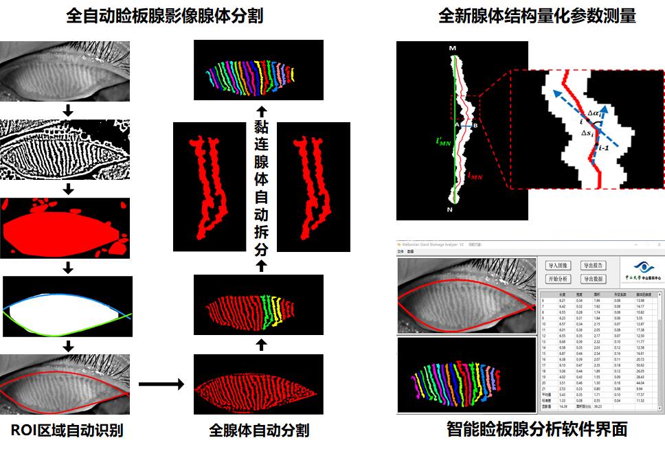
The fully automated algorithm was based on infrared image contrast enhancement and feature recognition technology for objective segmentation and multi-parametric quantitative analysis of meibography images. Besides of traditional parameters, such as glands area ratio (GA), to objectively quantify the gland diameter variations, new parameters such as deformation index (DI), the gland tortuosity (TI) as well as the glands signal index (SI) were proposed, offering more comprehensive information which is likely to be advantageous for detecting local and subtle changes of meibomian glands.
The principle and procedure of the automated Meibograhy analyser
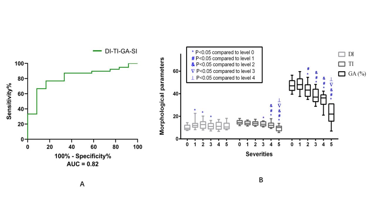
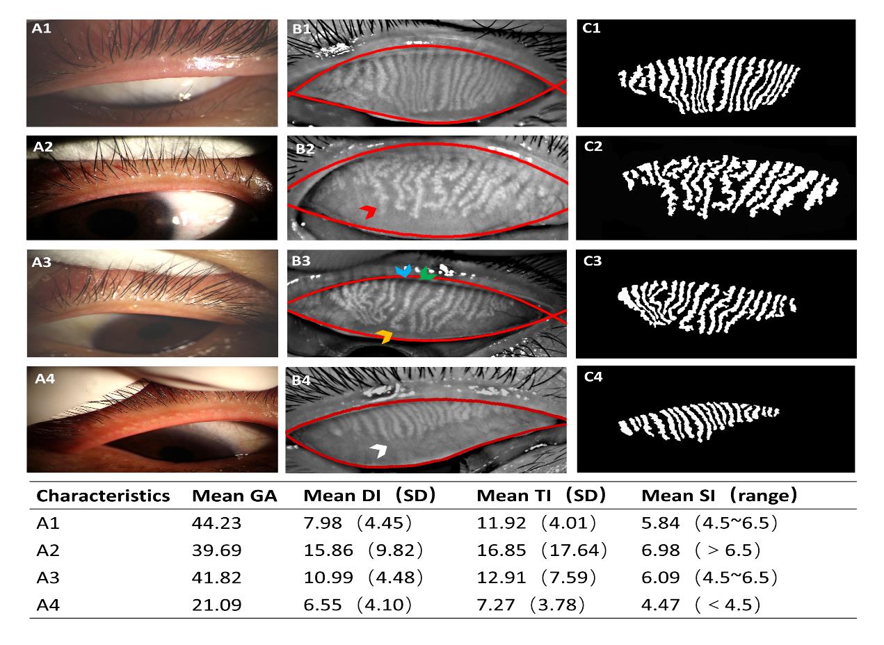
With the automated multiparametric quantitative analysis of MGs in meibography images, the real world research found that an increased DI (cross-sectionally uneven gland dilation), a decreased GA (greatly influenced by incomplete atrophy) and a decreased TI (decreased axial distortion) were associated significantly with the presence of MGD. A decline TI with a reduction of the GA can represent overall gland atrophy, which is different from gland dropout or shortened glands, as a complement sign of severe loss of acinar cells. The study also revealed that glands with a low-level SI showed a high risk for the presence of MGD. The combination of DI-TI-GA-SI showed an excellent diagnosis performance in identification of MGD patients from people with suspected DED (AUC=0.82), which will benefit to fast screen for MGD in dry eye clinics.
The analysis results of representative meibography images
Moreover, since the morphology and function of meibomian glands varies at different MGD stages, multi-parametric analysis offering more comprehensive information could help in discovering subtle changes of meibomian glands during MGD progression. The study found that morphological features including an increased DI and a slightly increased GA are common in the early stage of MGD, while a decreased GAtogether with a decreased TI are signs of severe MGD. The merge of morphological features (TI-DI-GA) showed excellent accuracy in distinguishing MGD severities (AUC 0.79~1.0), which provides a new standard for clinical MGD grading.

The diagnosis and grading performanceof MGD with quantitative parameters
At present, this intelligent meibomian glands analysis system has been commercialized, and installed to the Meibography imaging systems. The first batch of equipment equipped with the software has entered clinical application, providing a new choice for the early diagnosis and quantitative grading of MGD.

The technology transfer signing ceremony


