Prof. Yizhi Liu's team redefines the core functions of lens epithelial cells in the life cycle and proposes new concepts for clinical application
Resource: State Key Laboratory of Ophthalmology
Proofread by: Jiawei Wang
Edited by: Xianjing Wei
Reviewed by: Xiaoling Liang
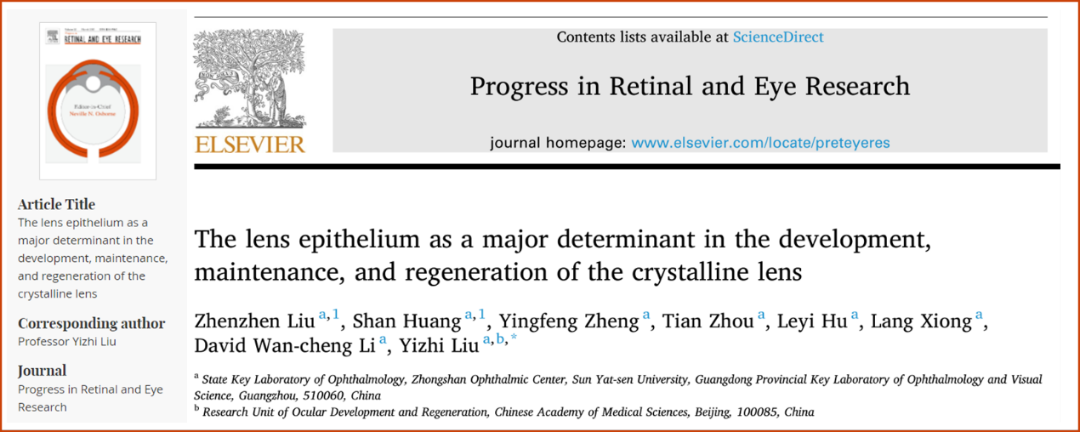
The lens is a clear, biconvex tissue in the eye that functions to focus light on the retina. Lens clouding, or cataract, is the world's leading cause of blindness. Surgical treatment with prostheses (Intraocular lens, IOLs) is commonly used for cataract, but the medical and socioeconomic costs are enormous. Keeping the human lens clear for life, or even reversing the clouding at an early stage, is the most ideal way to prevent and treat cataracts, and is the relentless pursuit of mankind. Intervention and regulation of the lens epithelium cells (LECs), which are the core cells of the lens, may be an effective way to achieve this goal. However, there is a lack of comprehensive understanding and utilization of their biological properties. Based on the long-term work of their team in this field, Professor Liu Yizhi from the Zhongshan Ophthalmic Center,Sun Yat-sen University discovered and elaborated the important roles of LECs in different processes of lens embryonic development, transparency maintenance and wound repair from the perspective of the whole life cycle, providing a theoretical basis for autologous cell-mediated tissue function maintenance and repair therapy, overturning the traditional concept of cataract clinical prevention and treatment, providing a future direction for research in this field. The article was published online on August 31, 2022 in the most influential journal of ophthalmology, Progress in Retinal and Eye Research, titled “The lens epithelium as a major determinant in the development, maintenance, and regeneration of the crystalline lens”.

Prof. Yizhi Liu's team looked back at the amazing process presented by LECs throughout the lens lifetime and illustrated the decisive role played by LECs in the three stages of embryonic development, homeostasis maintenance and postoperative regeneration.
During the embryonic stage, a monolayer of cells derived from the epidermal ectoderm form lens vesicle. Then, different parts of the lens are subject to different regulatory signals to form different parts of the three-dimensional structure of the lens. The posterior and equatorial LECs undergo key events such as cell cycle exit, crystallin expression, cell body elongation, cellular organelles degradation and nucleus removal to form primary and secondary lens fibers respectively. The anterior LECs differentiate and expand to form the epithelial layer, some of which retain the ability to differentiate into fiber cells. These events are influenced by genetic or environmental factors, Their abnormality will lead to abnormal lens development. Genetic screening using genome-wide genotyping and genome-wide association studies (GWAS), analysis of RNA and protein structures using X-ray crystallography and cryo-electron microscopy can help reveal how genetic defects impair protein function within the LECs and provide new insights into the molecular mechanisms underlying abnormal lens development.
After the lens matures, LECs under the anterior capsule and at equatorial regions maintain lens homeostasis through substance synthesis and exchange while slowly proliferating and differentiating. The potential difference between the cell membranes of LECs and lens fiber cells in different areas forms the lens microcirculation, which transports energy, oxygen and other substances. Lens microcirculation disorder causes cataract. Cholesterol synthesis disorder also causes lens clouding. Lanosterol is an important intermediate product of cholesterol synthesis. It can stabilize molecular chaperons and enhance proteasome activity to improve lens proteolysis and maintain or restore lens transparency.
After lens surgery, we can harness the biological properties of LEC proliferation and differentiation for clinical treatment. Incomplete differentiation and fibrosis of LECs produces cloudy tissue, while complete differentiation and orderly arrangement produces clear lens tissue. In the pseudophakic eyes, where the implanted prosthesis plays an optical role, the proliferation of LECs affects the function of IOLs. The LECs can be removed or inhibited using techniques such as the square-edge barrier effect of the IOL and fluid jet posterior capsule polishing technique. In infants and children, the size of the anterior and posterior capsulorhexis at cataract extraction can be improved to promote the formation of Soemmerring ring in the peripheral part of the aphakic eye and facilitate the implantation of IOL in the capsular bag at stage II. Through an innovative minimally invasive lens surgery, the contents of the cloudy lens can be removed while preserving the LECs, so as a clear lens with refractive power can be regenerated in situ to improve the prognosis of cataract in infants and children.
Maintaining lifelong lens transparency in human is the ultimate goal in cataract prevention and treatment. Investigating and exploiting the biological characteristics of LECs could be an effective way to achieve this goal.
Link to original article:
https://authors.elsevier.com/a/1fgRa4pAsjh4fM
