Abstract: Cavernous hemangioma is the most primary benign orbital tumor in adults, and majority of cases could be easily settled by surgical treatment. However, cavernous hemangioma lodged deep in the orbital apex remained a challenge because the surgery may pose a high risk of injury to the optic nerve and significant visual loss. This presentation would report a case of cavernous hemangioma located in orbital apex who presented superonasal and inferotemporal peripheral vision defect. The patient received fully transnasal endoscopic surgery, and a 2 cm × 1.5 cm tumor was successfully removed from the left orbital apex. The treatment results were satisfactory, with no after-effects and adverse reactions during follow-up. This case highlighted that transnasal endoscopic surgery is a promising technique for cavernous hemangiomas that are located deep in orbital apex. This approach provides direct pathway to tumor with limiting morbidity, maximal surgical field and ample illumination. The procedure represents a safe and less invasive management.
Abstract: Vogt-Koyanagi-Harada syndrome (VKH) is a bilateral granulomatous panuveitis associated with serous retinal detachments and vitritis, and can be associated with extraocular manifestations of meningismus, poliosis, vitiligo, hearing loss, and headaches. It is mediated by CD4+ T cells that target melanocytes in the eye, ear, meninges, and skin. It classically presents in 4 different phases: prodromal, uveitic, convalescent, and recurrent. There have been considerable advances in our understanding of the disease in recent years, and options for treatment have also expanded beyond systemic corticosteroids though these remain the mainstay of therapy in patients with VKH. This brief review will focus on updates in the diagnosis and treatment of VKH, specifically advances in imaging techniques including the use of optical coherence tomography angiography (OCTA) and enhanced depth imaging (EDI) optical coherence tomography (OCT). OCT parameters that are diagnostically predictive of acute VKH compared to other exudative maculopathies include the presence of subretinal membranous structures, a high retinal detachment, subretinal hyperreflective dots, and RPE folds. Evaluations of choroidal thickness using EDI-OCT demonstrate predominant involvement of the outer choroid in the acute inflammatory phase of VKH, consistent with histopathological analysis. OCTA may emerge as an alternative to fluorescein angiography (FA) and indocyanine angiography (ICGA) but is limited at this time due to its small field of view. While the mainstay of treatment of acute VKH continues to be systemic corticosteroids, biological response modifiers (BRMs) such as adalimumab and infliximab have been shown to be effective in the management of adult and pediatric VKH with one benefit being a faster onset of action compared to conventional immunosuppression. Literature Search: A literature search was done in PubMed using the words “Vogt Koyanagi Harada” “imaging” “diagnosis” “treatment” “therapy “posterior uveitis”.
Abstract: Uveitis can cause significant visual morbidity and often affects younger adults of working age. Anterior uveitis, or inflammation limited to the anterior chamber (AC), iris, and/or ciliary body comprises the majority of uveitis cases. Current clinical biomarkers and conventional grading scales for intraocular inflammation are mostly subjective and have only a moderate degree of interobserver reliability, and as such they have significant limitations when used in either clinical practice or research related to uveitis. In recent years, novel imaging techniques and applications have emerged that can supplement exam findings to detect subclinical disease, monitor quantitative biomarkers of disease progression or treatment effect, and provide overall a more nuanced understanding of disease entities. The first part of this review discusses automated algorithms for optical coherence tomography (OCT) image processing and analysis as a means to assess and describe intraocular inflammation with higher resolution than that afforded by conventional AC and vitreous cell ordinal grading scales. The second half of the review focuses on anterior segment OCT and OCT angiography (OCTA) in scleritis and iritis, especially with regards to their ability to directly image and characterize the pathologic structures and vasculature underlying these diseases. Finally, we briefly review experimental animal research with promising but more distant human clinical applications, including in vivo molecular microscopy of inflammatory markers and investigation of gold nanoparticles as a potential contrast agent in OCT imaging. Imaging modalities are discussed in the broader context of trends within the field of uveitis towards greater objectivity and quantifiable outcome measures and biomarkers.
Abstract: Optical coherence tomography angiography (OCTA) is a fast, non-invasive imaging modality that provides detailed information on retinal and choroidal vascular flow and macular structure. OCTA offers an accurate three-dimensional view of the individual retinal vascular plexuses and the choriocapillaris which facilitates the detection of the microvascular abnormalities in a variety of macular diseases. The perfusion indices (vessel density and flow index) are valuable parameters evaluated by OCTA that allow a quantitative interpretation of changes in the retinal vasculature that can reflect the severity of disease. Crystalline retinopathy encompasses a group of conditions whose distinctive feature is the presence of retinal crystals often located in the posterior pole. Select crystalline retinopathies also demonstrate retinal vascular abnormalities as well. Considering that the OCTA is a novel imaging modality and crystalline retinopathies are relatively rare conditions, there are currently few reports of OCTA findings associated with crystalline retinopathy. The advent of OCTA allows visualization of vascular and structural changes in crystalline retinopathies that are unique and cannot be appreciated on other imaging modalities, including fluorescein angiography (FA). This article reviews novel OCTA findings which provide new insights in the pathogenesis of crystalline retinopathies, including Bietti crystalline retinopathy, talc retinopathy, macular telangiectasia type 2, tamoxifen retinopathy, and Sj?gren-Larsson Syndrome maculopathy.
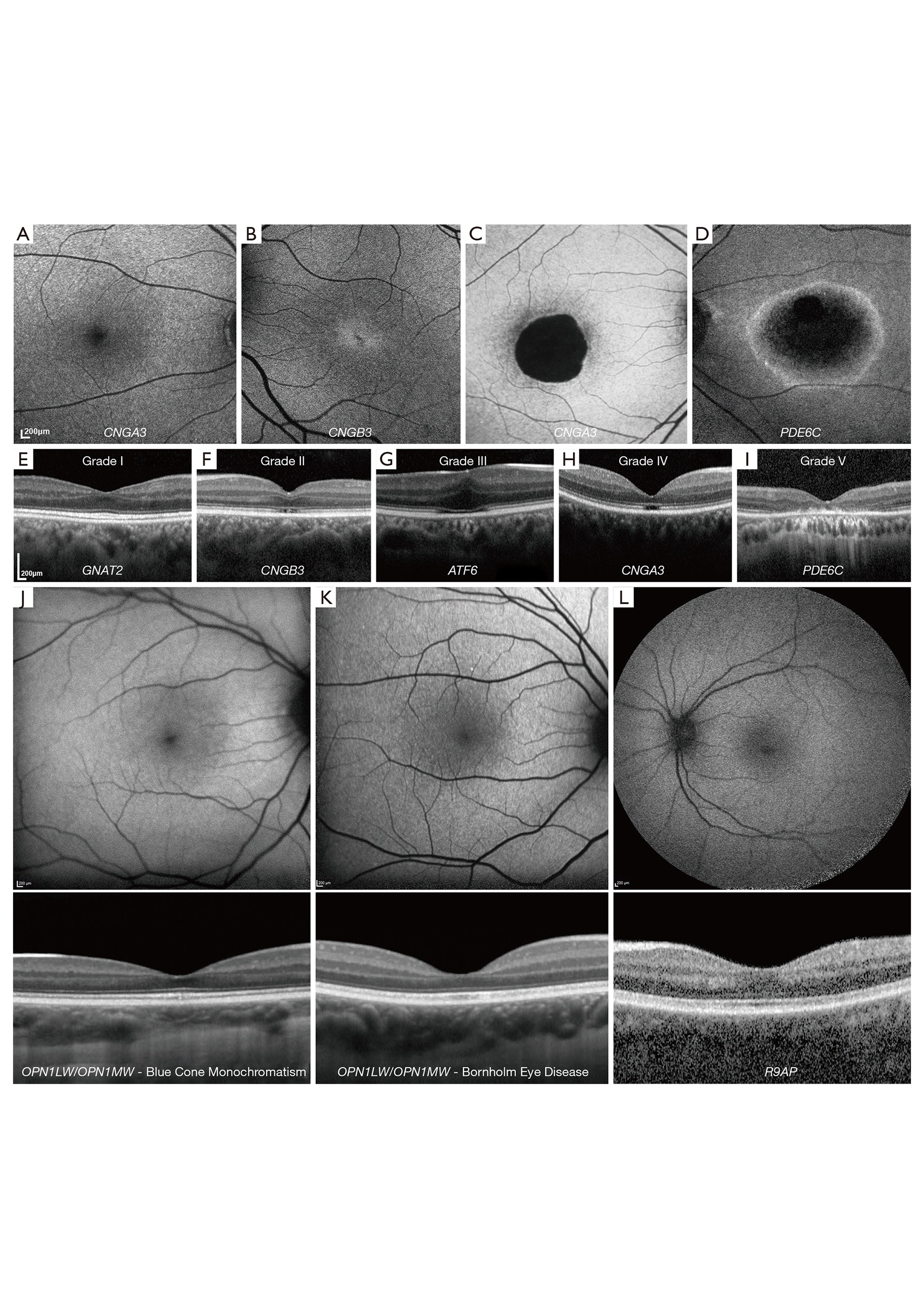
Abstract: Inherited retinal diseases (IRD) are a leading cause of blindness in the working age population. The advances in ocular genetics, retinal imaging and molecular biology, have conspired to create the ideal environment for establishing treatments for IRD, with the first approved gene therapy and the commencement of multiple therapy trials. The scope of this review is to familiarize clinicians and scientists with the current landscape of retinal imaging in IRD. Herein we present in a comprehensive and concise manner the imaging findings of: (I) macular dystrophies (MD) [Stargardt disease (ABCA4), X-linked retinoschisis (RS1), Best disease (BEST1), pattern dystrophy (PRPH2), Sorsby fundus dystrophy (TIMP3), and autosomal dominant drusen (EFEMP1)], (II) cone and cone-rod dystrophies (GUCA1A, PRPH2, ABCA4 and RPGR), (III) cone dysfunction syndromes [achromatopsia (CNGA3, CNGB3, PDE6C, PDE6H, GNAT2, ATF6], blue-cone monochromatism (OPN1LW/OPN1MW array), oligocone trichromacy, bradyopsia (RGS9/R9AP) and Bornholm eye disease (OPN1LW/OPN1MW), (IV) Leber congenital amaurosis (GUCY2D, CEP290, CRB1, RDH12, RPE65, TULP1, AIPL1 and NMNAT1), (V) rod-cone dystrophies [retinitis pigmentosa, enhanced S-Cone syndrome (NR2E3), Bietti crystalline corneoretinal dystrophy (CYP4V2)], (VI) rod dysfunction syndromes (congenital stationary night blindness, fundus albipunctatus (RDH5), Oguchi disease (SAG, GRK1), and (VII) chorioretinal dystrophies [choroideremia (CHM), gyrate atrophy (OAT)].
Abstract: An intestinal dysbiosis is connected to a number of inflammatory diseases through various mechanisms relating to its effect on immune cell function and differentiation. This is a review of the literature summarizing our current understanding of intestinal microbial contributions to non-infectious uveitis and strategies to target the intestinal microbiome to treat uveitis. Several groups have demonstrated an intestinal dysbiosis associated with certain types of non-infectious uveitis. Additionally, approaches to treat uveitis by modifying the intestinal microbiota, such as oral antibiotics or administration of oral short chain fatty acids (SCFAs), which are intestinal bacterial metabolites produced by fermentation of dietary fiber, can successfully treat uveitis in mouse models. This reduction in severity of ocular inflammation occurs via the following mechanisms: enhancement of regulatory T cells, decreasing intestinal permeability, and/or affecting T cell trafficking between the intestines and the spleen. Other strategies that are directed at the intestinal microbiota that might be effective to treat uveitis include dietary changes, probiotics, or fecal microbial transplantation. The commensal gut bacteria are influential in systemic and intestinal mucosal immunity and thus contribute to the development of extraintestinal inflammation like uveitis. Targeting the intestinal microbiome thus has the potential to be a successful strategy to treat non-infectious uveitis.
Abstract: Acute retinal necrosis (ARN) is a devastating syndrome characterized by panuveitis, retinal necrosis, and a high rate of retinal detachment that may result in poor visual outcomes if not promptly diagnosed and treated. ARN is most commonly caused by viruses within the herpesvirus family. Etiologies include varicella-zoster virus, herpes simplex virus, and cytomegalovirus, and may be promptly diagnosed by polymerase chain reaction testing of aqueous or vitreous fluid. The true incidence of ARN is not known due to its rarity; as a result, clinical treatment is often guided by retrospective case series, case reports, and expert opinion. Standard of care has evolved over time but currently includes a combination of systemic and intravitreal antiviral in conjunction with topical or oral steroids and surgical therapy as needed. Combination therapy may reduce the rate of severe vision loss and increase the rate of visual acuity gain, although further studies are needed in this area. In particular for patients with mild to moderate disease, combination therapy may reduce the rate of retinal detachment. Adjunctive therapies including oral corticosteroid and prophylactic laser barricade are incompletely studied, but corticosteroid in particular, may reduce inflammation, which also is involved in the severe disease pathogenesis observed in ARN. This review discusses the advances in diagnosis and treatment of ARN, including management with combination antiviral medication and surgical interventions.
Abstract: Autoimmune retinopathy (AIR) refers to both paraneoplastic and non-paraneoplastic forms of a rare, acquired retinal degeneration thought to be mediated by the production of antiretinal antibodies. However, the mechanisms underlying AIR pathogenesis are incompletely understood, and it remains a diagnosis of exclusion given the lack of definitive testing as well as its protean clinical presentation. This review summarizes the current literature on the epidemiology, diagnosis, and management of AIR, with a focus on non-paraneoplastic disease and the potential role of immunomodulatory therapy. A recent expert consensus statement on diagnosis and management of non-paraneoplastic AIR served as a framework for interpreting the limited data available, a process that was complicated by the small sample sizes, heterogeneity, and retrospective nature of these studies. Additional work is needed to characterize AIR patients on the basis of cytokine and immunogenetic profiling; to establish the pathogenicity of antiretinal antibodies; and to standardize treatment regimens as well as assessment of clinical outcomes.