一亿年进化出的晶状体有多重要?
How important is the lens evolved in 100 million years?
发布时间:2022-12-01 浏览:2988
谭源
导语:
“让科学研究走出象牙塔,用学术科普打破科学研究的神秘‘黑洞’”——本期视频的灵感来源为“内源性干细胞在晶状体再生修复中的应用”,以《眼科学报》2022年5月“封面论文”为基础进行创作。3分钟的时间里,我们用科普动画与真人讲解的形式为大家讲述“从自然界生物进化出晶状体,到研究者们在蝾螈为代表的低等两栖动物中观察到晶状体再生,再到在人类中实现功能性晶状体再生”的故事。
大约在46亿年前,我们的地球诞生了。过了几十亿年,大约在5亿年前的寒武纪,地球上开始出现了生命。寒武纪早期的时候,地球上的生物是没有眼睛的。大约在寒武纪晚期的时候,出现了具有感光功能的眼点,这些眼点由感光细胞组成,是最原始的眼睛,但眼点接收到的光线是杂乱的。
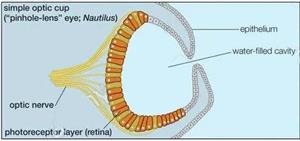
经过漫长的演化,一部分生物率先将感光细胞排列成像杯子一样的形状,此时可以判断光从哪个方向来,形成了更具竞争力的视觉器官。

随后生物向形成更典型的眼睛迈步,杯子的侧面形成一个针孔,这个小孔可以滤过杂乱的光线,位于眼睛底部的感光细胞逐渐感受到了物体的影像。
蜗牛等动物在此基础上进一步发育出来了球形的透明细胞团构成的晶状体,从而使眼睛能够聚焦。

泥盆纪时,眼球前段出现了可以变焦的晶状体,不同距离物体可以在视网膜上形成清晰物象。
Introduction:
"Let scientific research go out of the ivory tower and break the mysterious" black hole "of scientific research with academic science popularization" - The inspiration of this video is "the application of endogenous stem cells in lens regeneration and repair", and it is based on the "cover paper" of the Journal of Ophthalmology in May 2022. In 3 minutes, we told the story of "lens evolved from natural creatures, researchers observed lens regeneration in lower amphibians represented by salamanders, and then realized functional lens regeneration in humans" in the form of popular science animation and live explanation.

About 4.6 billion years ago, our earth was born. After several billion years, about 500 million years ago in the Cambrian period, life began to appear on the earth. In the early Cambrian, life on the earth had no eyes. Around the late Cambrian, eye spots with photosensitive function appeared. These eye spots are composed of photosensitive cells and are the most primitive eyes, but the light received by the eye spots is messy.
After a long evolution, some organisms took the lead in arranging photoreceptors into the shape of cups. At this time, we can judge the direction of light and form a more competitive visual organ.
Later, the creature moves to form a more typical eye. A pinhole is formed on the side of the cup. This pinhole can filter out the messy light. The photoreceptor cells at the bottom of the eye gradually feel the image of the object.
On this basis, snails and other animals further developed lenses composed of spherical transparent cell clusters, so that the eyes can focus.
During the Devonian period, lenses that can be zoomed appeared in the anterior segment of the eye, and objects at different distances can form clear images on the retina.

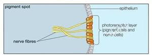
Nowadays, we can see objects clearly, and the lens plays an important role. When the external light enters the eyes, it can form a clear image on the retina after being refracted by the lens and other refractive media. The lens is originally transparent, but it may appear cloudy for various reasons. The opacification of the lens is called cataract, which can cause blurred vision, glare, ghosting, etc. The main treatment of cataract is to remove the cloudy lens in the eye and implant intraocular lens. However, for infants with cataract, traditional surgical treatment may lead to severe postoperative inflammatory reaction and many complications.
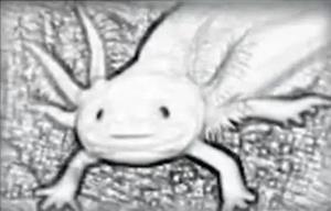
In 1895, scientists observed lens regeneration in lower amphibians represented by salamanders, which shows that lens can regenerate in nature. Scientists began to explore lens regeneration.
More than a hundred years later, scientists realized the preservation of stem cells and regenerative microenvironment derived from the lens body in mammalian models such as New Zealand rabbits and cynomolgus monkeys, and finally achieved functional lens regeneration in infant cataract patients. So far, scientists have achieved the goal of using endogenous stem cells to regenerate a new functional lens. From the appearance of lens, to the regeneration of amphibian lens, to the regeneration of human functional lens, what new surprises will we get from the research of lens in the future?
返回列表
|